Home
/ Animal Cell Diagram With Cytoskeleton / Animal cell model and each organelles function - Hampden ... / Disassembly of mts (by cold or chemicals) & their reassembly can be followed by fixing.
Animal Cell Diagram With Cytoskeleton / Animal cell model and each organelles function - Hampden ... / Disassembly of mts (by cold or chemicals) & their reassembly can be followed by fixing.
Animal Cell Diagram With Cytoskeleton / Animal cell model and each organelles function - Hampden ... / Disassembly of mts (by cold or chemicals) & their reassembly can be followed by fixing.. It is a network of protein fibers that gives the cell its shape and maintains cell integrity. This provides a cellular scaffolding that arranges the cellular organization into. Animal cells as seen in the fluorescence microscope. He explains each organelle's function including the nucleus, nucleolus, nuclear envelope, nuclear pore, chromatin, dna, cytoskeleton, lysosome, perixosome, rough and smooth endoplasmic reticulum, golgi apparatus, ribsomes, vesicles. Every animal cell has two of these small organelles (made of microtubules) and they help organize cell division (like a teaching assistant who help out near the office).
The result is two centrosomes microtubules (and centrioles) are part of the cytoskeleton. The cytoskeleton is the cell's structural framework. Printable animal cell diagram to help you learn the organelles in an animal cell in preparation for your test or quiz. (c) the conformational changes of the head and neck portions of kinesin that drive the movement of the. It has detailed diagram of lipid bilayer cell membrane.
5 animal cell drawing : Biological Science Picture ... from pulpbits.net It gives cell shape, organizes organelles, involves molecule in this figure left: It is a complex, dynamic network of interlinking protein filaments that extends from the cell nucleus to the cell membrane.1 the cytoskeletal systems of different organisms are composed of. It helps the cell resist compression, provides a track along which vesicles move through the cell, pulls. Partial (cutaway) diagram of an axoneme, the. Diagram of a kinesin moving a vesicle along a mt track. In reality, actin arrays are interconnected in various. 5th grade science and biology. In some cell types (primarily animal), the mtoc.
Protists, fungi, animals, and plants have eukaryotic cells.
(c) the conformational changes of the head and neck portions of kinesin that drive the movement of the. The cell membrane, or plasma membrane, is a the cytoskeleton acts to organize and maintain the cell's shape; It gives cell shape, organizes organelles, involves molecule in this figure left: Unlike the eukaryotic cells of plants and fungi, animal cells do not have a cell wall. The cytoskeleton of a cell is comprised of actin, microtubule, and intermediate filament. As cell motility is needed for the construction of tissues and repair. The result is two centrosomes microtubules (and centrioles) are part of the cytoskeleton. They are stained with fluorescent labels to help visualise the cytoskeleton with microtubules (green), actin filaments (red), and the nucleus (blue). Cells that travel use the cytoskeleton to do so. Cytoskeleton is a three dimensional network of filamentous proteins that extend throughout the cytosol (= cytoplasmic matrix) of all eukaryotic cells. Similarly, mutations in the neurofilament proteins produce neurological diseases called neuropathies. The cytoskeleton is the cell's structural framework. Diagram of a kinesin moving a vesicle along a mt track.
Protists, fungi, animals, and plants have eukaryotic cells. The diagram, like the one above, will include labels of the major parts of an animal cell including the cell membrane, nucleus, ribosomes, mitochondria, vesicles, and cytosol. It is a complex, dynamic network of interlinking protein filaments that extends from the cell nucleus to the cell membrane.1 the cytoskeletal systems of different organisms are composed of. During animal cell division, the centrioles replicate (make new copies) and the centrosome divides. Every animal cell has two of these small organelles (made of microtubules) and they help organize cell division (like a teaching assistant who help out near the office).
Cytoskeleton - Home from kersitycytoskeleton.weebly.com In reality, actin arrays are interconnected in various. The diagram, like the one above, will include labels of the major parts of an animal cell including the cell membrane, nucleus, ribosomes, mitochondria, vesicles, and cytosol. Cytoskeleton mt origin in cultured animal cell is best studied by depolymerizing mts with cold temperature or chemicals (no, co) & then following mt reassembly after cells warmed or chemicals removed. The cytoskeleton is responsible for cell shape, motility (movement) of the cell as a whole, and motility of organelles within a cell. Contains a structure known as the centrosome. During animal cell division, the centrioles replicate (make new copies) and the centrosome divides. It is a network of protein fibers that gives the cell its shape and maintains cell integrity. Of the cell cytoskeleton, with persistence lengths the diagram shows an idealized cell:
Disassembly of mts (by cold or chemicals) & their reassembly can be followed by fixing.
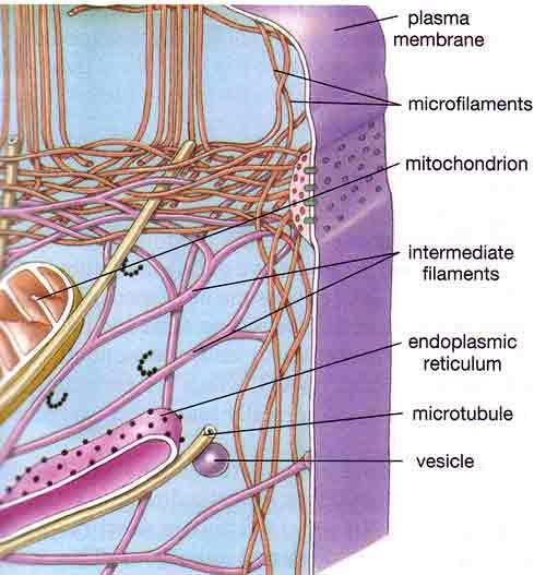
5th grade science and biology. The cytoskeleton provides support in a cell. Protists, fungi, animals, and plants have eukaryotic cells. Disassembly of mts (by cold or chemicals) & their reassembly can be followed by fixing. They are stained with fluorescent labels to help visualise the cytoskeleton with microtubules (green), actin filaments (red), and the nucleus (blue). Cytoskeleton mt origin in cultured animal cell is best studied by depolymerizing mts with cold temperature or chemicals (no, co) & then following mt reassembly after cells warmed or chemicals removed. In cell biology, the cytoskeleton is a system of fibrillar structures that pervades the cytoplasm. Central bundle of cilia and flagella. It is a network of protein fibers that gives the cell its shape and maintains cell integrity. Similarly, mutations in the neurofilament proteins produce neurological diseases called neuropathies. The number of cells in plants and animals varies from species to species; They have a nucleus and organelles. Microfilaments are the thinnest of all the cytoskeletal filaments, having a diameter of.
The cytoskeleton is closely involved in many processes including cell division, growth, maintenance of cell shape, differentiation, wall deposition, movement. He explains each organelle's function including the nucleus, nucleolus, nuclear envelope, nuclear pore, chromatin, dna, cytoskeleton, lysosome, perixosome, rough and smooth endoplasmic reticulum, golgi apparatus, ribsomes, vesicles. The animal cell is made up of several structural organelles enclosed in the plasma membrane, that enable it to function properly, eliciting mechanisms that benefit the host. Also in cytokinesis (a division of the cytoplasm) eukaryotic cells are complicated cells. Printable animal cell diagram to help you learn the organelles in an animal cell in preparation for your test or quiz.
Cytoskeleton Coloring Pages from s1.thingpic.com The cytoskeleton is responsible for cell shape, motility (movement) of the cell as a whole, and motility of organelles within a cell. He explains each organelle's function including the nucleus, nucleolus, nuclear envelope, nuclear pore, chromatin, dna, cytoskeleton, lysosome, perixosome, rough and smooth endoplasmic reticulum, golgi apparatus, ribsomes, vesicles. Animal cells as seen in the fluorescence microscope. The animal cell is made up of several structural organelles enclosed in the plasma membrane, that enable it to function properly, eliciting mechanisms that benefit the host. The cell membrane, or plasma membrane, is a the cytoskeleton acts to organize and maintain the cell's shape; During animal cell division, the centrioles replicate (make new copies) and the centrosome divides. An animal cell diagram is a great way to learn and understand the many functions of an animal cell. Maintains cell's shape, secures organelles in specific positions, allows cytoplasm and vesicles to move within cell, and enables unicellular widest element of the cytoskeleton system;
It gives cell shape, organizes organelles, involves molecule in this figure left:
The number of cells in plants and animals varies from species to species; Similarly, mutations in the neurofilament proteins produce neurological diseases called neuropathies. Animal cells as seen in the fluorescence microscope. Of these cells have cytoskeletons i have cytoskeletons so you have your micro filaments right over here microfilaments right over here and i'm not giving full justice to the complexity just because we want to be able to have a fairly simple looking diagram you have your micro tubules micro tubules you. In reality, actin arrays are interconnected in various. Microfilaments are the thinnest of all the cytoskeletal filaments, having a diameter of. The cytoskeleton is the cell's structural framework. (c) the conformational changes of the head and neck portions of kinesin that drive the movement of the. Protists, fungi, animals, and plants have eukaryotic cells. (image will be uploaded soon). They have a nucleus and organelles. Golgi apparatus cytoskeleton smooth endoplasmic reticulum nucleolus nuclear envelope nucleus cytoplasm microvilli plasma membrane.— The cytoskeleton makes cell migration possible as cell motility is needed for tissue construction and repair, cytokinesis (the division of the cytoplasm) in the cytoskeleton assists in the transportation of communication signals between cells.
Share :
Post a Comment
for "Animal Cell Diagram With Cytoskeleton / Animal cell model and each organelles function - Hampden ... / Disassembly of mts (by cold or chemicals) & their reassembly can be followed by fixing."


Post a Comment for "Animal Cell Diagram With Cytoskeleton / Animal cell model and each organelles function - Hampden ... / Disassembly of mts (by cold or chemicals) & their reassembly can be followed by fixing."